An Introduction to Mesoporous Silica Nanoparticles
Mesoporous silica nanoparticles (MSNs) are silica nanoparticles with pores that range in diameter from 2 to 50 nm and have an overall diameter below 1 µm. Their range of pore sizes is consistent with the IUPAC definition of mesoporous1 and make them ideal materials for applications ranging from catalysis and environmental chemical removal to biomedicine. Here at Fortis Life Sciences, we bring the synthetic expertise required to tailor the design of MSNs to target an intended application with the utmost control.
Design Characteristics of Mesoporous Silica Nanoparticles
In addition to their biocompatibility, key advantages of MSNs are associated with their high surface area and facile surface tunability. Often described as "nanosponges," MSNs can adsorb large amounts of molecules dissolved in solution thanks to this high surface area; in fact, 5 grams of mesoporous silica nanoparticles possess the same surface area as an American football field. This highly ordered porosity can be controlled during the synthesis of MSNs.
Tunable Pore Sizes for Size-Selective Adsorption
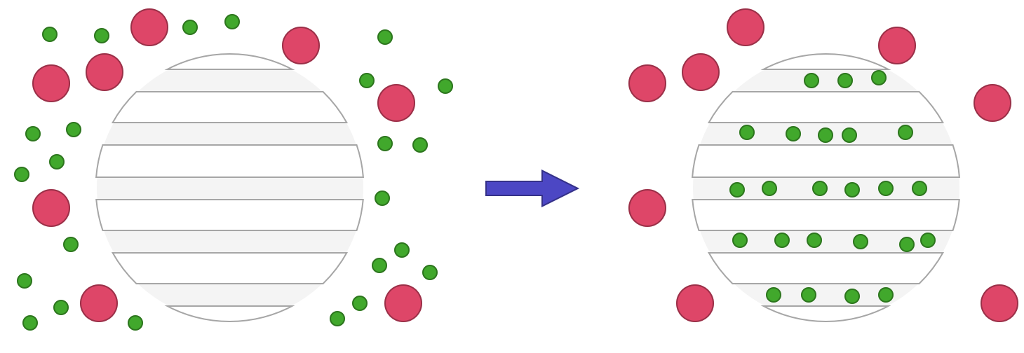
MSNs can be made to have very narrow pore size distributions, yielding nanoparticles that will selectively adsorb molecules of a specific size. The figure below illustrates that only molecules within a specific size range (small green circles) will enter the MSN cavities, while other larger molecules (large red circles) will be excluded. This feature is useful for molecule separation by size exclusion.
Tunable Surface Chemistry for Functionally-Selective Adsorption
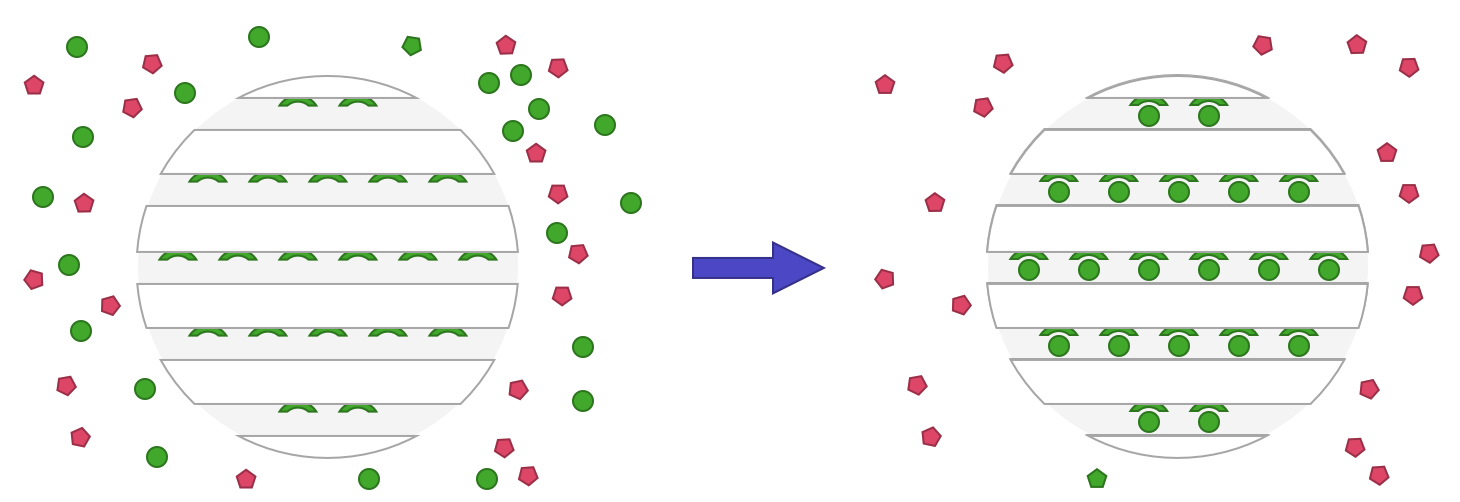
Thanks to the versatility of the silica, it’s possible to modify the surfaces of MSNs with specific functional groups that will interact selectively with certain categories of molecules. One can imagine coating the pores with a positive surface in order to attract negatively charged molecules, or using a hydrophobic surface to attract more hydrophobic molecules. The figure below shows an MSN in which the pores are modified to allow molecules with a specific surface functionality (represented as green circles) to adsorb inside the particles, whereas others (red pentagons) are excluded. This type of selective adsorption has numerous uses in hydrophobic drug encapsulation, environmental chemical removal, and various other applications.
Variable Pore Structures for Controlled Release
The organizational structure of the pores can also be modulated (MCM-41, MCM-48, radial, cubic, wormlike, etc.), as represented in the following figure. The use of one type over the others will depend on the application, as the pore structure controls the release/leakage of molecules loaded inside the pores.
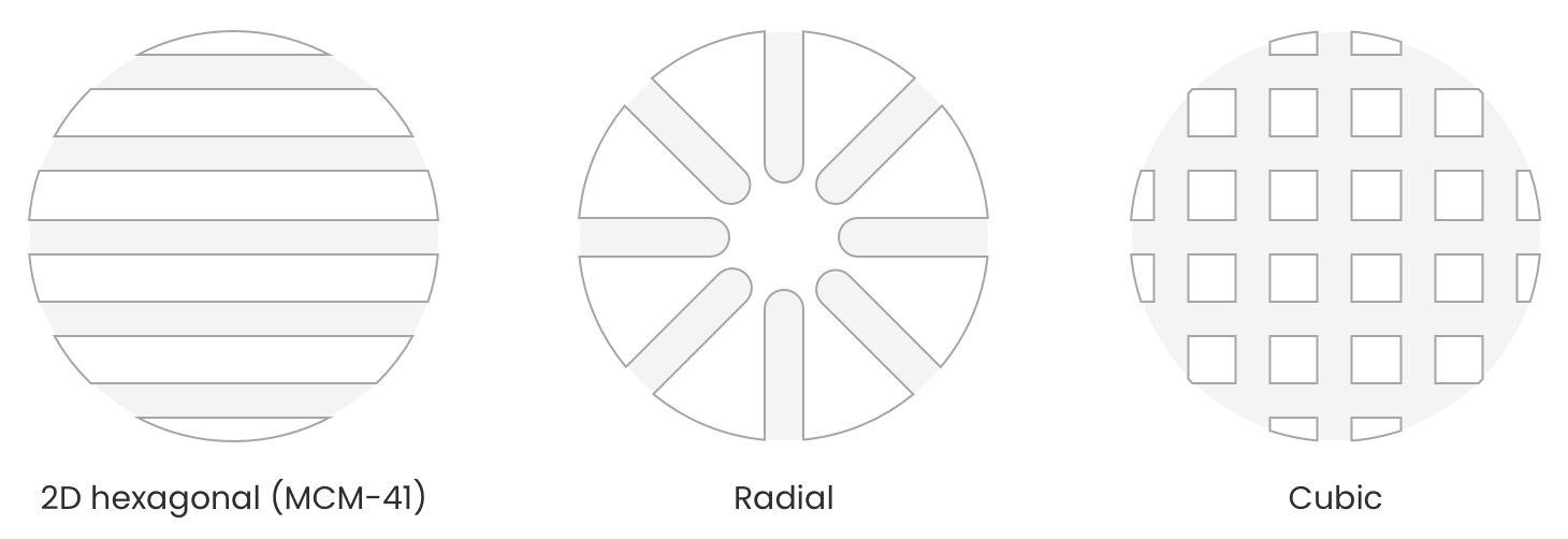
For example, MSNs with the hexagonal pore arrangement can release cargo from both ends of a specific channel whereas in the cubic arrangement, molecules can travel freely inside the intricate network and be released from any pore outlet. In the radial pore arrangement, the molecule can only access a single pore and thus must exit through the pore it entered, making this particular system less prone to premature leaking (in the case of drug encapsulation).
Applications of Mesoporous Silica Nanoparticles
In general, we can look to the unique structural properties of MSNs to justify how these materials are used. Here you’ll find a brief overview of how these properties are useful to biomedicine and catalysis.
Nanomedicine
Over the past 15 years, the myriad benefits of MSNs have majorly impacted the drug delivery field, evidenced by the rising number of scientific articles involving their use as drug carriers.2–4 Their high surface area, easy surface modification, and biocompatibility make them the material of choice for targeted delivery of active pharmaceutical ingredients for cancer therapy and other treatments.2 The high surface area of MSNs allows them to carry larger amounts of the desired drug than through conventional methods (e.g. polymers, liposomes, and vesicles), and with the size-selective capabilities discussed previously. Moreover, the easy surface modification and biocompatibility makes them useful for targeted delivery of active pharmaceutical ingredients with different chemistries such as molecules with hydrophilic and/or hydrophobic properties. The surface functionalization allows for the potential targeting of specific tissues or cells, or the increase of the enhanced permeability and retention (EPR) effect by evading the reticuloendothelial system (RES). Although mesoporous silica is generally robust, MSNs can be engineered to be degradable to facilitate controlled release of their contents.5
Catalysis
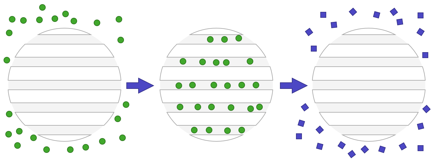
The easily adjustable surface chemistry gives MSNs a unique affinity for loading catalysts onto their high surface areas. MSNs (MCM41-type, in particular) have shown shape selective capabilities of the substrate due to the narrow pore size distribution. This has been observed in the epoxidation of bulky alkene substrates using titanocene-grafted-MSN structures.6 Grafting can be achieved through activating the surface silanols (or other surface chemistry) by displacing the ligands of the catalyst species and attaching to the inorganic core. Inorganic catalysts can also be integrated into the structure of the MSNs to increase catalytic efficiency, for instance titanium can be integrated into a MCM41-structured zeolite for oxidation.7 The cartoon below illustrates the use of MSNs for catalysis, where the analyte (green circles) penetrates the pores of the particles where the catalysis reaction happens, and a new product is then formed and released (purple squares).
Furthermore, surface functionalized MSNs have been shown to exhibit catalytic capability on their own using general acid/base chemistry without the use of transition metals. As an example, it has been demonstrated using bi-functionalized MSNs with both an acid and base group to perform carbonyl activation for Aldol, Henry, and cyanosilylation reactions. Examination of the reaction rates based on the varying ratios of acid/base functionalization suggest the MSNs take part in the reaction mechanism.8
A key benefit of MSNs is the ease of centrifugation instead of requiring chemical separation to isolate product, which decreases the resources needed and makes the process greener. The mechanical separation won’t damage the catalysts or mesoporous framework so the catalytic material can be regenerated or recycled easily.
Synthesis & Purification of Mesoporous Silica Nanoparticles
Compared to the synthesis of regular silica nanoparticles that generally follow the Stöber process, MSNs are synthesized in a water-based solution in the presence of a base catalyst and a pore forming agent more widely known as a surfactant. Surfactants are molecules that present the particularity to have a hydrophobic tail (alkyl chain) and a hydrophilic head (charged group, such as a quaternary amine for example). As these surfactants are added to a water-based solution, they will coordinate to form micelles with increasing concentration in order to stabilize the hydrophobic tails as shown below.
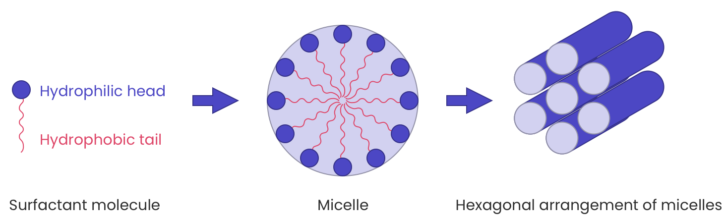
During the early stage of the hydrolysis and condensation of the silica precursor, oligomeric forms of silica appear. Like the final material, these oligomers possess silanol groups (Si-OH) that are deprotonated under the basic conditions of the reaction, forming negatively charged oligomers that can in turn condense on the surface of the positively charged micelles. As the reaction proceeds, the oligomers grow larger around the micelles ultimately forming a hybrid organic/inorganic silica network templated by surfactant molecules. After the reaction, the removal of the template will create the porosity that defines mesoporous silica nanoparticles.
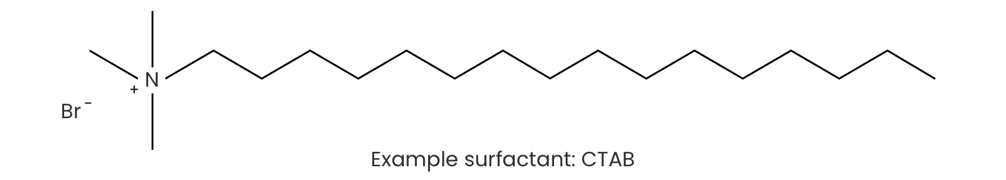
Purification: The Importance of Removing CTAB for Biological Applications
After synthesis, CTAB must be removed for practical use of MSNs. This is important for three main reasons:
Pore accessibility
The presence of the surfactant inside the pores will reduce the capability of the particles to encapsulate any active pharmaceutical ingredient (API) by decreasing the pore volume.
Cytotoxicity
Most of the common surfactants used to synthesize mesoporous silica nanoparticles are harmful to the body. The widely used CTAB molecule is able to interact with the phospholipids constituting the cell membrane which could lead to cell death at high concentrations. As a result it is crucial to ensure total removal of these pore forming agents prior to biomedical application, especially injections.
Pore surface modification
Removal of the surfactant molecules from the pores will make them accessible to further modification such as amination, thiolation, or hydrophobization. Doing so makes it possible to have the same functionality throughout the entire silica surface (inside the pores and outside of the particles), or to have two distinct types of functionality which will bring different capabilities to each spatial region. An example would be the loading of a negatively charged molecule inside the pores (after amine modification of the inner surface) while having the outside of the particle functionalized with thiol or any other functional group for targeted drug delivery.
CTAB Removal Methods
Solvent Extraction
The removal of the CTAB molecules from the particles by solvent extraction can be done using either a solvent mixture of concentrated hydrochloric acid and ethanol or an ethanolic solution of ammonium nitrate. Both methods require the use of heat (around 60 °C) for at least 1 hour. They are generally performed twice to ensure that all the CTAB molecules have been removed from the pores. In general, the smaller and more tortuous the pores, the more extraction the sample requires.
Calcination
One of the most common techniques for surfactant removal is calcination. This process involves heating dried samples of mesoporous silica in an oven set above 500 °C for five hours or more. At the elevated temperature, the surfactants that reside inside the pores decompose, leaving the desired mesoporous silica framework intact. This technique allows us to process a large amount of mesoporous silica but runs the risk of damaging the pore structure.
Pros & Cons
While the calcination method is able to completely free the pores from any organic molecule and surfactants, it cannot be used if the surface of the particle is already modified (with amines, thiols, or carboxylic groups) for the same reason. Moreover the calcination method can promote irreversible aggregation due to dehydration and crosslinking between particles.
The ammonium nitrate or hydrochloric acid treatments are more compatible with the presence of functionalities on the surface of the particles, but may lead to incomplete surfactant removal if the pores are too small.
Custom Mesoporous Silica Nanoparticles at Fortis Life Sciences
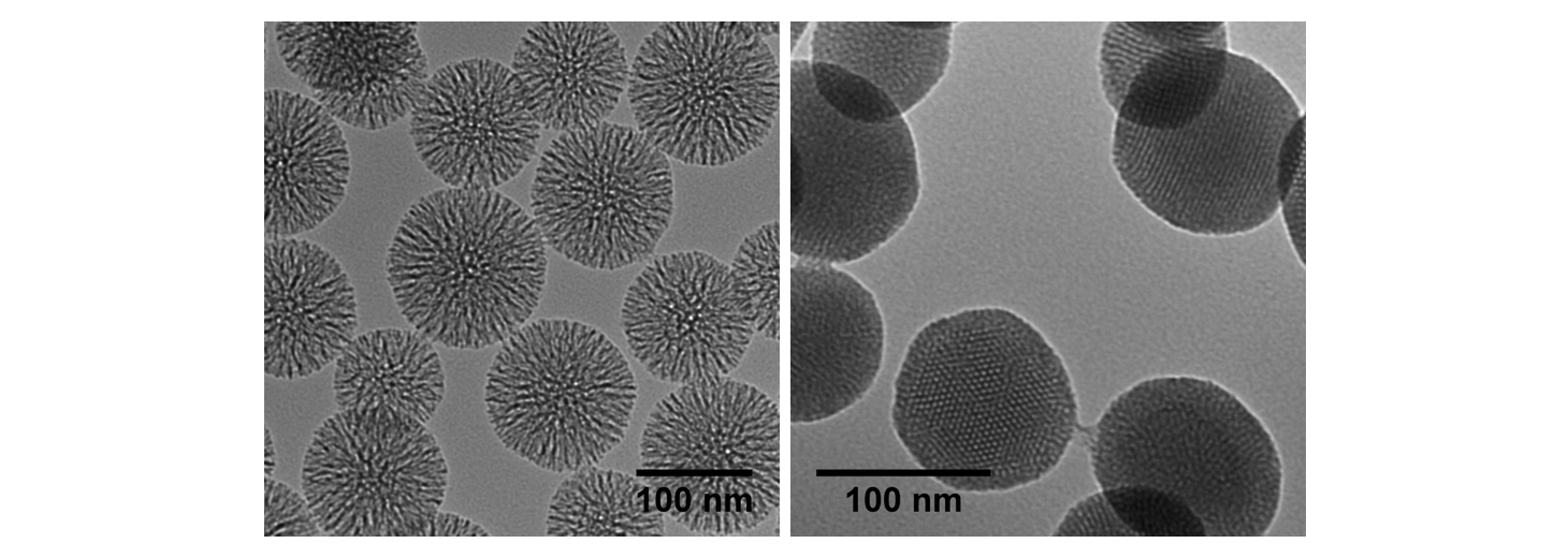
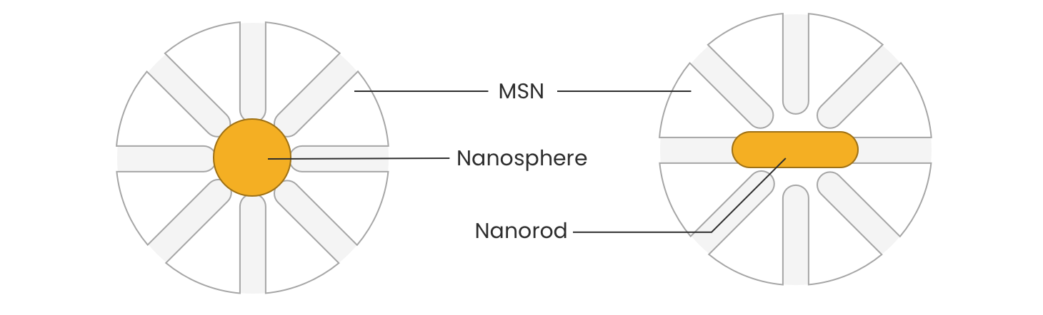
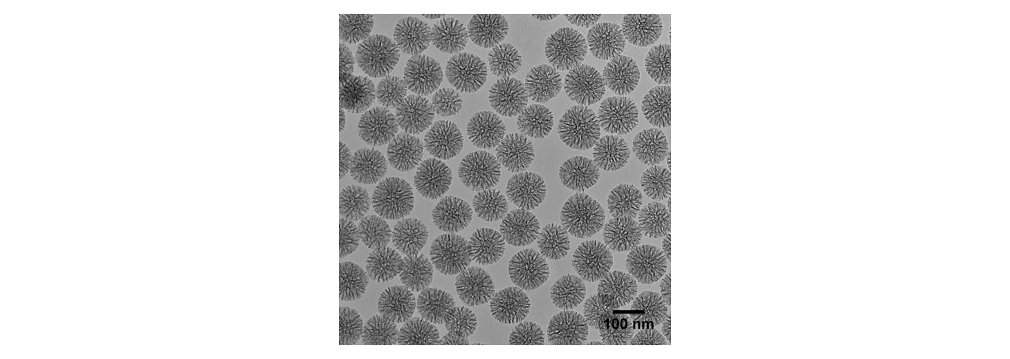
At Fortis Life Sciences, we can prepare MSNs of various sizes (from 50 to several hundreds of nanometers in diameter) and with different pore arrangements such as the widely used MCM-41 (hexagonal pore) and radial types of MSNs. The following TEM images demonstrate the distinction between radial (left) and hexagonal (right) pore geometries. The 100 nm MCM-41 pore particles on the right are available here at Fortis Life Sciences as a standard product. The radial pore particles can be ordered on a custom basis.
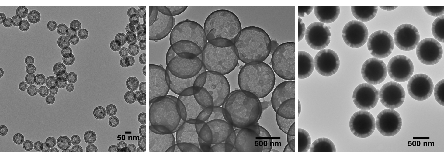
It is also possible to design and synthesize nanoparticles with various structures by modulating the porosity; such nanoparticles can have a hollow interior and a mesoporous silica shell, or a larger hollow interior and a thin microporous silica shell. At Fortis Life Sciences we can also prepare particles with a microporous core and a thick mesoporous shell; the possibilities are almost infinite and limited only by the imagination. The TEM images below show MSNs with variable porosity in different spatial regions of the particles. These images show 50 nm MSNs with highly porous interiors and a denser shell/coating (left), 500 nm hollow silica nanoparticles (HSNs; middle), and 500 nm solid silica nanoparticles with mesoporous shells (right).
Mesoporous-Silica Shelled Nanoparticles
The presence of the surfactants (e.g. CTAB) in the synthesis of MSNs conveniently enables the incorporation of a core of different material types at the center of the nanoparticle. Cores made up of other inorganic materials and even other types of silica can be suspended in these aqueous surfactant solutions for subsequent shell deposition and growth.
For these core-shell syntheses, the inorganic nanoparticles are generally stabilized directly by a surfactant on their surface. The presence of the surfactants provides the steric stability needed for the inorganic nanoparticles to be dispersed in aqueous solutions. The extra layer of CTAB on the surface of the inorganic particles can act as an anchor point for the solubilized silicate species (obtained after hydrolysis of the silica precursor), leading to the growth of silica around the inorganic nanoparticles. This technique has been used to prepare core-shell nanoparticles composed of silver, gold, iron oxide or quantum dot cores surrounded by a mesoporous silica shell. The image below shows TEM of 50 nm gold nanoparticles shelled with mesoporous silica.

Such materials combine both the advantages of the mesoporous silica (biocompatibility, drug delivery platform) with the unique properties of the core (plasmonics, imaging, photothermal, magnetic, etc.) and are widely used in the relatively new research field of theranostics. It can also be applied to different particle morphologies to make a wide variety of interesting heterostructures (see image below).
Large-Pore Mesoporous Silica Nanoparticles
Since 2010, there has been increased interest and research done on synthesizing mesoporous silica with large dendritic pores ranging from 5–30 nm in diameter.9 There are multiple pore expanding agents that are in current use today ranging from ring structures such as cyclohexane and trimethyl benzene, to chain structures such as octadecene.10, 11 There have also been successful syntheses using less harsh agents like sodium salicylate and sodium trifluoroacetate for an anion enhanced expansion.12 The TEM image below shows one example of the large pore MSNs we fabricate here at Fortis Life Sciences.
Additional Resources
Featuring nanoComposix Mesoporous Silica Nanoparticles
- Zhao, Q.; Wu, B.; Shang, Y; Huang, X.; Dong, H.; Liu, H.; Chen, W.; Gui, R.; Li, J. Development of a Nano-drug Delivery System Based on Mesoporous Silica and its Anti-lymphoma Activity. Appl. Nanosci. 2020. https://doi.org/10.1007/s13204-020-01465-0.
References Cited
- Rouquerol, J.; Avnir, D.; Fairbridge, C. W.; Everett, D. H.; Haynes, J. M.; Pernicone, N.; Ramsay, J. D. F.; Sing, K. S. W.; Unger, K. K. Recommendations for the Characterization of Porous Solids (Technical Report). Pure Appl. Chem. 2009, 66 (8), 1739–1758. doi.org/10.1351/pac199466081739.
- Li, Z.; Barnes, J. C.; Bosoy, A.; Stoddart, J. F.; Zink, J. I. Mesoporous Silica Nanoparticles in Biomedical Applications. Chem. Soc. Rev. 2012, 41 (7), 2590–2605. doi.org/10.1039/C1CS15246G.
- Slowing, I. I.; Vivero-Escoto, J. L.; Wu, C.-W.; Lin, V. S.-Y. Mesoporous Silica Nanoparticles as Controlled Release Drug Delivery and Gene Transfection Carriers. Adv. Drug Deliv. Rev. 2008, 60 (11), 1278–1288. doi.org/10.1016/j.addr.2008.03.012.
- Argyo, C.; Weiss, V.; Bräuchle, C.; Bein, T. Multifunctional Mesoporous Silica Nanoparticles as a Universal Platform for Drug Delivery. Chem. Mater. 2014, 26 (1), 435–451. doi.org/10.1021/cm402592t.
- Hao, X.; Hu, X.; Zhang, C.; Chen, S.; Li, Z.; Yang, X.; Liu, H.; Jia, G.; Liu, D.; Ge, K.; et al. Hybrid Mesoporous Silica-Based Drug Carrier Nanostructures with Improved Degradability by Hydroxyapatite. ACS Nano 2015, 9 (10), 9614–9625. doi.org/10.1021/nn507485j.
- Maschmeyer, T.; Rey, F.; Sankar, G.; Thomas, J. M. Heterogeneous Catalysts Obtained by Grafting Metallocene Complexes onto Mesoporous Silica. Nature 1995, 378 (6553), 159–162. doi.org/10.1038/378159a0.
- Corma, A.; Navarro, M. T.; Pariente, J. P. Synthesis of an Ultralarge Pore Titanium Silicate Isomorphous to MCM-41 and Its Application as a Catalyst for Selective Oxidation of Hydrocarbons. J. Chem. Soc. Chem. Commun. 1994, No. 2, 147–148. doi.org/10.1039/C39940000147.
- Huh, S.; Chen, H.-T.; Wiench, J. W.; Pruski, M.; Lin, V. S.-Y. Cooperative Catalysis by General Acid and Base Bifunctionalized Mesoporous Silica Nanospheres. Angew. Chem. Int. Ed. 2005, 44 (12), 1826–1830. doi.org/10.1002/anie.200462424.
- Polshettiwar, V.; Cha, D.; Zhang, X.; Basset, J. M. High-Surface-Area Silica Nanospheres (KCC-1) with a Fibrous Morphology. Angew. Chem. Int. Ed. 2010, 49 (50), 9652–9656. doi.org/10.1002/anie.201003451.
- Chen, F.; Goel, S.; Valdovinos, H. F.; Luo, H.; Hernandez, R.; Barnhart, T. E.; Cai, W. In Vivo Integrity and Biological Fate of Chelator-Free Zirconium-89-Labeled Mesoporous Silica Nanoparticles. ACS Nano 2015, 9 (8), 7950–7959. doi.org/10.1021/acsnano.5b00526.
- Shen, D.; Yang, J.; Li, X.; Zhou, L.; Zhang, R.; Li, W.; Chen, L.; Wang, R.; Zhang, F.; Zhao, D. Biphase Stratification Approach to Three-Dimensional Dendritic Biodegradable Mesoporous Silica Nanospheres. Nano Lett. 2014, 14 (2), 923–932. doi.org/10.1021/nl404316v.
- Wang, Y.; Song, H.; Yang, Y.; Liu, Y.; Tang, J.; Yu, C. Kinetically Controlled Dendritic Mesoporous Silica Nanoparticles: From Dahlia- to Pomegranate-like Structures by Micelle Filling. Chem. Mater. 2018, 30 (16), 5770–5776. doi.org/10.1021/acs.chemmater.8b02712.