Table of contents:
- Storage and Handling Tips
- Verifying Nanoparticle Stability
- Agglomeration After Exposure to Media
- Dissolution
Storage and Handling Tips
Nanoparticles can be extremely sensitive to their environment and require careful storage and handling. Here are some tips for maintaining nanoparticle stability:
- Minimize the time nanoparticles are exposed to light, heat, or air by storing in the dark, keeping material in the fridge, and recapping bottles immediately after use.
- Aliquot by pouring instead of inserting a pipet tip directly into the bottle. If required, extensively rinse the pipet tip with high purity water before immersing.
- Shake bottle or container vigorously before use. Metal nanoparticles will settle over a period of days but should easily disperse without sonication by shaking
Verifying Nanoparticle Stability
Careful storage and handling procedures must be followed in order to ensure that nanoparticles remain stable during storage. Depending on the nanoparticle's physical and chemical properties, they may be sensitive to light, temperature, and air exposure. To ensure the reproducibility and relevance of experiments, testing should be performed before nanoparticles are used in experiments. Depending on the equipment available in each laboratory, the following tests can be performed:
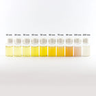
- Visual Identification: Silver and gold nanoparticles have a unique color in solution due to the plasmon resonance effect. The color of solution should be observed when the nanoparticles arrive and any changes to the color over time is an indication that the nanoparticles have changed. If there are any larger particulates floating in solution this is indicative of agglomeration and further testing should be performed.
- UV-Visible Spectroscopy: Measurement of the absorption of a the nanoparticle solution as a function of wavelength (typically, 400 - 1000 nm), is one of the best methods of tracking nanoparticle stability. Once the nanoparticles arrive, a baseline UV-visible spectrum should be acquired and compared to future measurements.
- Dynamic Light Scattering (DLS): DLS monitors the nanoparticle hydrodynamic diameter as a function of time and is sensitive to nanoparticle agglomeration. Establishing a base reading and then monitoring the DLS diameter as a function of time will provide data on the stability of the nanoparticle system.
- Transmission Electron Microscopy (TEM): TEM can be used to confirm the size and shape distributions provided by nanoComposix. In addition, a phenomenon known as Ostwald Ripening occurs with small nanoparticles. This effect can increase the size distribution of a nanoparticle sample even if no agglomeration occurs. The basis of this effect is that the dissolution rate of metal ions from the surface of smaller nanoparticles is greater than from the surface of larger nanoparticles. When the ions reabsorb onto the surface of the nanoparticles, the larger nanoparticles grow at the expense of the smaller nanoparticles resulting in increased size distributions. This is primarily an issue for high concentration of small nanoparticle solutions (< 10 nm diameter). TEM measurements will determine if there has been any change to the size distribution via Ostwald Ripening.
Agglomeration After Exposure to Media
When nanoparticles are exposed to high salt solutions such as those present in most biologically compatible media, agglomeration can occur. Depending on the nanoparticle material, size, and surface, the agglomeration can happen instantaneously or over a period of days. Once the particles agglomerate, they behave like much larger particles and can have rapid settling rates which complicates the measurement of dose.
It is critical to understand the rate of agglomeration of nanoparticles in the target system. Due to the presence of cells or other biological or environmental material, it is often not possible to directly monitor the agglomeration state once the nanoparticles are delivered. Therefore, it is highly recommended that stability studies be performed in a particulate free medium that is a close mimic to the target environment. Monitoring the UV-Visible spectrum or DLS hydrodynamic diameter over time will provide information on the rate of agglomeration. If the rate is rapid, then additional care must be taken to ensure that the timing associated with dosing events is carefully managed in order to make sure that the nanoparticles are in a reproducible agglomeration state when introduced into the experiment.
Dissolution
For nanoparticles such as silver, ion release from the surface is an important component of the toxicity of the nanoparticles. Dissolution rates are a function of the nanoparticle surface, size, temperature, and air exposure (primary due to oxygen and sulfur). Dissolution rates are measured by isolating the nanoparticles from the solution via filtration or centrifugation and then measuring the residual silver ion concentration. Since metal ions are known to have an effect on biota, it is important to understand the contribution of toxicity due to the nanoparticles and due to the free ions in solution.