Introduction
For applications requiring the highest uniformity in size and shape, nanoComposix Ultra Uniform Gold Nanospheres have a nearly perfect spherical shape, smooth surfaces, and the lowest coefficients of variation available anywhere (CV < 5%).

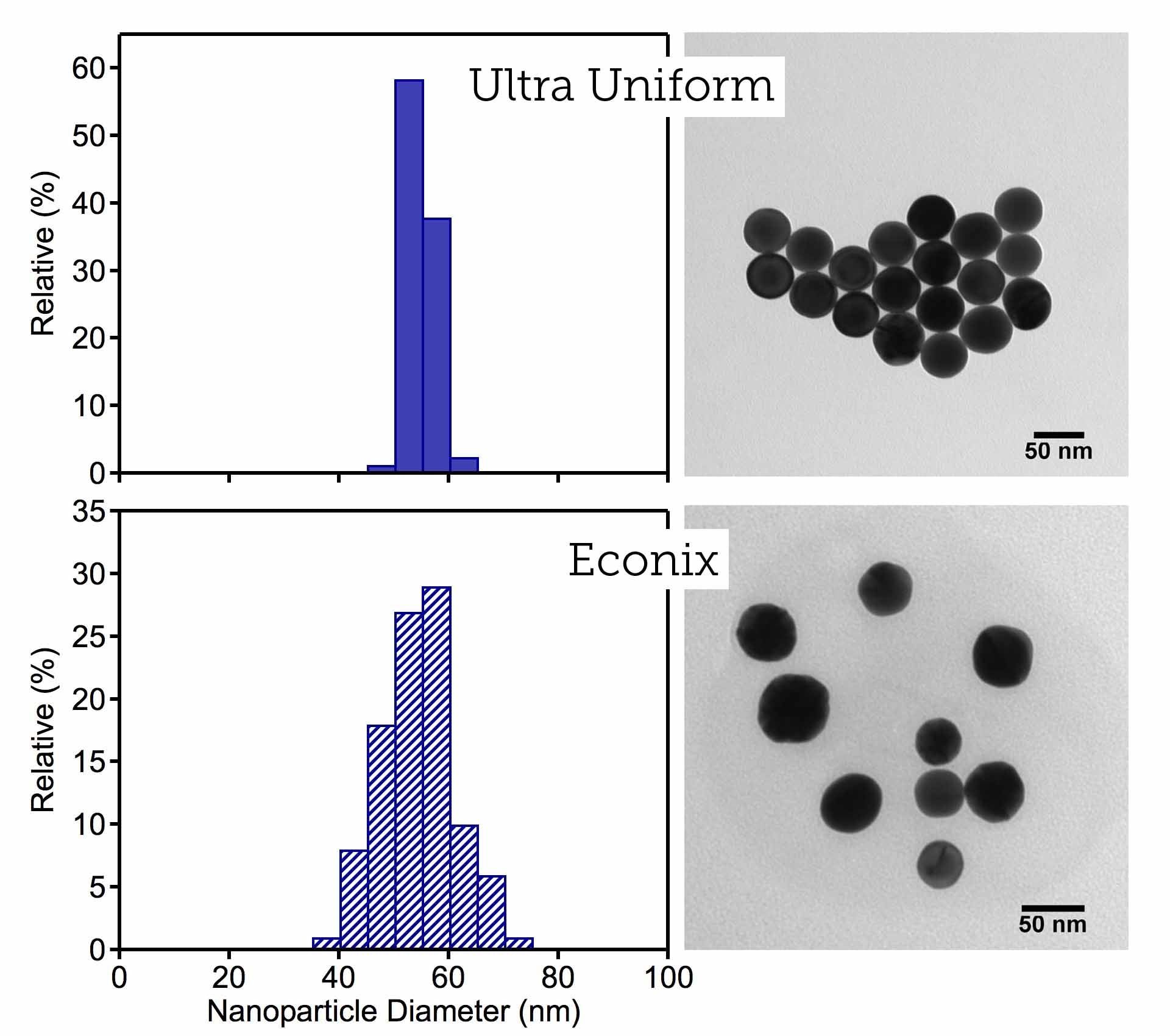
These particles are used as reference materials and for imaging techniques that take advantage of their exceptional uniformity. The histogram and images below compare size distributions of 50 nm Gold Nanospheres from our Ultra Uniform product line with those of our Econix product line (discontinued), as measured by transmission electron microscopy (TEM). The histogram and images highlight the narrow distribution of sizes in an ensemble of Ultra Uniform particles.
Ultra Uniform Gold Nanospheres are available as standard products with mean diameters of 10 nm, 20 nm, 30 nm, 50 nm and 100 nm. Other sizes up to 200 nm are available upon request. The particles are supplied at a concentration of 0.05 mg/mL in a sodium citrate solution and functionalized with a carboxyl surface to promote long term stability and to facilitate coupling to an antibody or other biomolecules using EDC/NHS binding chemistry.
Optical Properties
Ultra Uniform Gold Nanospheres exhibit greater optical density per unit mass (OD/mg) than our other gold nanoparticles at the same diameter, and their narrow size distributions give rise to extremely narrow optical extinction peaks. The normalized optical spectra below compare the width of the extinction bands for 100 nm gold nanoparticles from our Ultra Uniform and NanoXact product lines. A narrower band shape indicates a more homogeneous ensemble of nanoparticles in the Ultra Uniform materials and results in enhanced color purity.

Due to their highly uniform size and shape, Ultra Uniform Gold Nanospheres scatter a single color of light under dark field microscope imaging. The high purity of their light scattering signatures makes these nanoparticles ideal scattering labels for imaging and building blocks for plasmonic nanostructures and devices. Shown below are dark field images of the scattered light from 50 nm (left) and 100 nm (right) Ultra Uniform Gold Nanospheres.

Applications
Biological Labels
Ultra Uniform Gold Nanospheres benefit from exceptional spectral selectivity and color purity with respect to particle size. Because the color is invariant from particle to particle within a single batch, multiple labels can be used in a single image where different size selections of gold nanoparticles can be conjugated with different antibodies to help demarcate unique biological targets.
Reference Materials for Electron Microscopy
 Ultra Uniform Gold Nanospheres coated with electron-transparent polystyrene facilitates self-assembly of nanoparticles into evenly spaced arrays of exceptionally uniform particles. These properties, combined with high image contrast, large interparticle spacings, ordered crystalline structure, and controllable size make them ideal reference materials for transmission electron microscopy (TEM). Compared to other reference materials that may be useful only within a narrow range of magnification, Ultra Uniform Gold Nanospheres offer the unique ability to serve as a calibration tool over a wide range of magnification levels. The high image contrast and ordered arrangement of particles allows application of automated tools to analyze images and size the nanoparticles. For more information, check out our white paper about using Ultra Uniform Gold Nanospheres for TEM calibration.
Particle Number Standards
Ultra Uniform Gold Nanospheres are used as particle number standards for the calibration of single-particle inductively coupled plasma mass spectrometry (ICP-MS) instruments, light scattering instruments, and particle counting instruments. Because they are highly crystalline and non-porous, these nanoparticles can be used to quantify porosity single particle ICP-MS methods.
Probe for Functional Pore Sizes of Ultrafiltration Membranes
Ultra Uniform Gold Nanospheres have a demonstrated ability to validate the functional pore sizes of commercial ultrafiltration membranes. A recent study executed a gold nanoparticle retention test and compared the size and concentration of nanoparticles in the suspensions before and after passing them through membranes. The functional sizes of the pores can be determined with extraordinary accuracy because the sizes of the gold nanoparticles are carefully measured, extremely uniform, and do not distort under pressurized test conditions. This technique is a useful counterpart to common membrane pore size characterization methods like gas-liquid displacement porosimetry (GLDP) and liquid-liquid displacement porosimetry (LLDP). It also provides an alternative means of measurement where very small pore sizes inhibit effective LLDP measurements.
Nanoantennas for Sensing
Gold Nanospheres have a demonstrated ability to serve as nanoantennas in plasmonic waveguides. A recent study4 incorporated nanoComposix Ultra Uniform Gold Nanospheres into a silver nanowire complex to act as nanoantennas in a chirality sensing structure. When exposed to incident light, the Ultra Uniform Gold Nanospheres propagated surface plasmon polaritons (SPPs) along the silver nanowire structure and enabled the detection of circular dichroism with sensitivity as good as 1 mdeg of ellipticity.
Additional Resources
Featuring nanoComposix ultra uniform gold nanospheres:
- Chan, Q.; Entezarian, M.; Zhou, J.; Osterloh, R.; Huang, Q.; Ellefson, M.; Mader, B.; Liu, Y.; Swierczek, M. Gold Nanoparticle Mixture Retention Test with Single Particle Detection: A Fast and Sensitive Probe for Functional Pore Sizes of Ultrafiltration Membranes J. Membrane Sci. 2020, 599, 117822.
- Keri, A.; Sápi, A.; Ungor, D.; Sebők, D.; Csapó, E.; Kónya, Z.; Galbács, G. Porosity Determination of Nano- and Sub-micron Particles by Single Particle Inductively Coupled Plasma Mass Spectrometry J. Anal. Atom. Spectrom. 2020, 35, 1139–1147.
- Payne, L. M.; Zilli, A.; Wang, Y.; Langbein, W.; Borri, P. Quantitative High-Throughput Optical Sizing of Individual Colloidal Nanoparticles by Wide-field Imaging Extinction Microscopy – The ‘Long Shadow’ Effect Proc. SPIE 2019, 10892.
- Rothe, M.; Zhao, Y.; Müller, J.; Kewes, G.; Koch, C. T.; Lu, Y.; Benson, O. Self-Assembly of Plasmonic Nanoantenna–Waveguide Structures for Subdiffractional Chiral Sensing ACS Nano 2021, 15 (1), 351–361.