Plasmonic nanoparticles are discrete metallic nanoparticles that have unique optical properties, and are increasingly being incorporated into commercial products and technologies. These technologies, which span fields ranging from photovoltaics to biological and chemical sensors, take advantage of the extraordinary efficiency of gold and silver plasmonic nanoparticles at absorbing and scattering light. Additionally, unlike most dyes and pigments, plasmonic nanoparticles have a color that depends on their size and shape and can be tuned to optimize performance for individual applications without changing the chemical composition of the material.


Figure 1: (Left) Gold and silver nanoparticles of varying sizes and shapes. From left to right: 80 nm silver nanospheres, 20 nm silver nanospheres, 40 nm gold nanospheres, 12 nm gold nanospheres, 200 nm silver nanoplates, 120 nm silver nanoplates, 60 nm silver nanoplates. (Right) Plasmonic nanoparticles can simultaneously have large absorption and scattering cross sections. Here the particles absorb green and red light, causing transmitted light to appear blue, and scatter red light, causing scattered light to appear red.
nanoComposix has developed a number of technologies which take advantage of plasmonic and photonic nanomaterials. These technologies include ultrabright surface enhanced Raman scattering (SERS) tags for multiplexed and multiparameter cytometry, surface enhanced fluorescence (SEF) nanotags for ultrasensitive detection of biomolecules, and as a new class of photothermal therapeutic materials. Additionally, we are developing technologies to improve the performance of photovoltaic cells and photonic waveguides.
For more information about nanoComposix materials for plasmonics and nanophotonics technologies, explore our Nanomaterials for Optical Engineering page or contact us.
More Information
- What is a Surface Plasmon?
- Applications
- Detailed Description of Plasmonic Nanoparticle Optical Properties
- References
What is a Surface Plasmon?
The remarkable optical properties of plasmonic materials occurs because the conduction electrons on the nanoparticle surface undergo a collective oscillation when excited by light at specific wavelengths (Figure 2, left). This oscillation, which is known as a surface plasmon resonance (SPR), results in the unusually strong scattering and absorption of light. In fact, plasmonic nanoparticles can have optical cross sections up to 10 times larger than their physical cross sections, allowing individual nanoparticles to be imaged using dark field microscopy (Figure 2, middle, right). Interested in learning more? Please see our the Plasmonics Tutorial in our Knowledge Base.
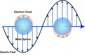


Figure 2: (Left) Schematic of surface plasmon resonance where the free conduction electrons in the metal nanoparticle are driven into oscillation due to strong coupling with incident light. (Middle) Dark field microscopy image of 50 nm gold nanoparticles. (Right) Dark field microscopy image of 60 nm silver nanoparticles.
Applications
Plasmonic nanoparticles have numerous applications including:
- Diagnostic Applications: Gold and silver nanospheres are used as reagents in numerous assays (including lateral flow assays) where the nanoparticles are used for qualitative or quantitative detection.
- Biosensing and Chemical Sensing Applications: Plasmonic nanoparticles including gold and silver nanospheres and silver nanoplates can be used to create surface-enhanced Raman scattering (SERS) tags for highly multiplexed biosensing1-3, and surface enhanced fluorescence (SEF, sometimes called metal enhanced fluorescence, MEF) tags for ultrasensitive detection4. Particles can also be engineered to detect ultralow concentrations of chemical agents by enhancing the intrinsic Raman scattering signal by up to 14 orders of magnitude.
- Optical Applications/Light Harvesting: Gold and silver nanospheres, silver nanoplates, and silver nanowires are used as nanoscale optical antennas to improve light harvesting in solar cells and other devices5.
- Therapeutics: Plasmonic nanoparticles are used to thermally ablate tumors where the extremely high absorption efficiency of the nanoparticles results in rapid particle heating that can selectively destroy targeted cancerous cells.
Detailed Description of Plasmonic Nanoparticle Optical Properties
Silver Nanospheres: The optical properties of silver nanospheres are a function of the nanoparticle diameter. As the diameter increases, the peak extinction (scattering + absorption) shifts to longer wavelengths and broadens, and the nanoparticle albedo (a ratio of scattering to total extinction) increases (Figure 3). At diameters greater than 80 nm, a second peak becomes visible at shorter wavelengths than the primary peak. This secondary peak is due to a quadrupole resonance that has a different electron oscillation pattern than the primary dipole resonance.


Figure 3: (Left) Extinction (the sum of scattering and absorption) spectra of nanoComposix silver nanoparticles with diameters ranging from 10 - 100 nm at mass concentrations of 0.02 mg/mL. 1 mg/mL nanoparticles have optical densities that are 50 times larger. (Right) Plot of silver nanosphere albedo (a ratio of scattering to total extinction) as a function of nanoparticle diameter.
Silver Nanoplates: The optical properties of silver nanoplates is a function of the aspect ratio (length:width), with plates with larger aspect ratios having peaks at longer wavelengths. By precisely controlling the plate diameter and thickness, the nanoplate’s optical resonance can be tuned to peak at specific wavelengths in the visible and near-IR spectral regions (Figure 4). This allows plates to be tuned to interact with specific laser lines, including 532 nm, 632.8 nm, 660 nm, 785 nm, 808 nm, and 1064 nm lasers.


Figure 4: (Left) Extinction spectra of silver nanoplates with varying aspect ratios. (Right) TEM image of silver nanoplates.
Gold Nanospheres: The optical properties of gold nanospheres is a function of the nanoparticle diameter. As the diameter increases, the peak plasmon resonance shifts to longer wavelengths and broadens, and the nanoparticle albedo (a ratio of scattering to total extinction) increases (Figure 5). Additionally, as the particles get larger, the particle scattering peak moves to longer wavelengths than the absorption peak (this can be observed by modeling different sized gold nanospheres using our Online Mie Theory Simulator).


Figure 5: (Left) Extinction (the sum of scattering and absorption) spectra of nanoComposix gold nanoparticles with diameters ranging from 10 - 100 nm at mass concentrations of 0.05 mg/mL. 1 mg/mL nanoComposix gold nanoparticles have optical densities that are 20-times larger. (Right) Plot of gold nanosphere albedo (a ratio of scattering to total extinction) as a function of nanoparticle diameter.
References
- D.S. Sebba, D.A. Watson, J.P. Nolan. High Throughput Single Nanoparticle Spectroscopy. ACS Nano, 2009, 3 (6) 1477-1484.
- R.T. Hill, J.J. Mock, D.S. Sebba, S.J. Oldenburg, S.Y. Chen, A.A. Lazarides, A. Chilkoti, D.R. Smith. Leveraging Nanoscale Plasmonic Modes to Achieve Reproducible Enhancement of Light. Nano Letters, 2010, 10 (10) 4150-4154.
- Willets, K.A. and R.P. Van Duyne, Localized Surface Plasmon Resonance Spectroscopy and Sensing. Annual Review of Physical Chemistry, 2007, 58 (1) 267-297.
- J.R. Lakowicz, C.D. Geddes, I. Gryczynski, J. Malicka, Z. Zhang, R. Badugu, J. Huang. Advances in Surface Enhanced Fluorescence. Journal of Fluorescence, 2004, 14 (4) 425-441.
- A.P. Kulkarni, K.M. Noone, K. Munechika, S.R. Guyer, D.S. Ginger. Plasmon-Enhanced Charge Carrier Generation in Organic Photovoltaic Films Using Silver Nanoprisms. Nano Letters, 2010, 10 (4) 1501-1505.
- D. P. O'Neal, L. R. Hirsch, N. J. Halas, J. D. Payne, J. L. West. Photo-thermal Tumor Ablation in Mice using Near Infrared-Absorbing Nanoparticles. Journal of Cancer Letters, 2004, 209, 171-176.