
Endotoxin contamination is an important consideration when nanoparticles are used in biological, toxicological, or pharmaceutical research. Even low levels of bacterial endotoxin can stimulate inflammatory and immune responses that may be incorrectly attributed to the nanoparticle itself.
Nanoparticles can also interfere with endotoxin assays through optical effects, aggregation, surface interactions, or other matrix effects. Reliable analysis therefore requires both an appropriate detection method and controls that demonstrate the assay can accurately detect endotoxin in the presence of the nanoparticle formulation.
Need endotoxin data for a nanoparticle sample?
Our endotoxin testing service uses a kinetic turbidimetric method adapted from USP <85> and Nanotechnology Characterization Laboratory methods for nanoparticle samples.
On this page
- What Endotoxin Is and Why It Matters
- Endotoxin Test Methods
- Kinetic Turbidimetric LAL Analysis
- Why Nanoparticles Interfere with Endotoxin Assays
- Sample Preparation and Dilution
- Inhibition and Enhancement Controls
- Interpreting Endotoxin Results
- Troubleshooting Nanoparticle Endotoxin Analysis
What Is Endotoxin and Why Does It Matter?
Endotoxins are lipopolysaccharides (LPS) associated with the outer membrane of gram-negative bacteria. LPS can be released as bacteria grow, shed membrane material, or undergo cell lysis.
The biologically active portion of LPS, lipid A, can activate innate immune signaling and trigger inflammatory responses. Depending on the exposure and biological system, endotoxin can contribute to cytokine release, fever, hypotension, and other responses.
This makes endotoxin an important experimental control in:
- Nanotoxicology studies
- Cell-based assays
- Immunological studies
- Preclinical nanomedicine development
- Parenteral pharmaceutical and medical-device development
Endotoxin is a pyrogen, but the terms are not interchangeable. Endotoxins are bacterial pyrogens derived from gram-negative bacteria, while other biological or chemical substances can also produce pyrogenic responses.
Why Endotoxin Is Reported in EU/mL
Endotoxin results are usually reported in endotoxin units per milliliter (EU/mL). An endotoxin unit represents biological activity relative to a reference endotoxin standard.
EU should not be converted to endotoxin mass using a universal conversion factor. The biological activity per unit mass can vary with endotoxin source, structure, preparation, and reference material, which is why biological activity is reported instead of simply reporting a mass concentration.
Common Bacterial Endotoxin Test Methods
Several bacterial endotoxin test formats are available. Traditional methods use Limulus Amebocyte Lysate (LAL), while newer methods can use recombinant proteins involved in the same endotoxin-responsive cascade.
| Method | Detection Principle | Key Considerations for Nanoparticles |
|---|---|---|
| Gel-clot LAL | Formation of a visible clot in response to endotoxin | Simple but relatively low-resolution and susceptible to sample interference. |
| Chromogenic LAL | Color formation following activation of the LAL cascade | Nanoparticles that absorb or scatter near the detection wavelength can interfere with the optical signal. |
| Kinetic turbidimetric LAL | Increase in turbidity as the endotoxin-dependent clotting reaction proceeds | Useful for many nanoparticle formulations, provided baseline optical density and aggregation are controlled. |
| Kinetic chromogenic LAL | Rate of chromogenic signal development | Can provide sensitive quantitative measurements but remains susceptible to optical and matrix interference. |
| Recombinant Factor C / recombinant cascade | Recombinant endotoxin-responsive proteins generate fluorescent or chromogenic signals | Provides a non-animal-derived alternative, but product-specific method suitability and nanoparticle interference still need to be demonstrated. |
USP <85> describes established bacterial endotoxin test methods, while USP <86> provides additional techniques using recombinant Factor C or recombinant cascade reagents. Method selection should consider the sample matrix, potential optical interference, regulatory requirements, and demonstrated suitability for the specific formulation.
Kinetic Turbidimetric LAL Analysis for Nanoparticles
nanoComposix currently uses a kinetic turbidimetric LAL method adapted from USP <85> and the National Cancer Institute's Nanotechnology Characterization Laboratory (NCL) protocols for nanoparticle samples.
In the kinetic turbidimetric assay, endotoxin activates an enzymatic cascade in the LAL reagent that progressively increases solution turbidity. The instrument monitors optical density at 660 nm as a function of time.
The time required for the signal to reach a defined optical-density threshold is referred to as the onset time. Samples containing higher endotoxin concentrations generally reach the threshold more quickly.

Example kinetic turbidimetric assay data. Onset time is compared with a calibration curve to determine endotoxin concentration.
Standard Curve and Quantification
Known endotoxin standards are analyzed to establish the relationship between endotoxin concentration and onset time. The measured sample response is then compared with this calibration curve.
For the NCL kinetic turbidimetric method, the standard curve requires at least three calibration standards and a correlation coefficient of at least 0.980. Replicate precision and quality controls are also evaluated before sample results are accepted.
The usable analytical range depends on the endotoxin reagent, instrument, standard curve, sample dilution, and matrix. Results outside the validated calibration range should not simply be extrapolated without appropriate justification.
Why Nanoparticles Can Interfere with Endotoxin Assays
Nanoparticle formulations can interfere with endotoxin measurements in ways that do not commonly occur with simple molecular solutions. Interference can affect the optical readout, the enzymatic assay chemistry, or the availability of endotoxin to the assay reagent.
Optical Interference
Gold, silver, and other nanoparticles can absorb and scatter strongly at wavelengths used by photometric endotoxin assays.
Chromogenic assays commonly detect color development near 405 nm. Silver nanoparticles can exhibit strong extinction in this region, making it difficult to distinguish the assay signal from the nanoparticle background.
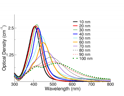
Silver nanoparticle extinction spectra overlap wavelengths used by some chromogenic endotoxin assays, creating the potential for optical interference.
High Baseline Optical Density
Kinetic turbidimetric analysis measures optical density at 660 nm. Large, concentrated, strongly scattering, or aggregated nanoparticles can already produce significant extinction at this wavelength before the LAL reaction begins.
A high starting optical density reduces the available dynamic range and can interfere with detection of the turbidity generated by the assay.
Aggregation During the Assay
The LAL reagent changes the chemical environment surrounding the nanoparticle. In some formulations, this can destabilize the dispersion and cause particle aggregation during the measurement.
Aggregation increases light scattering and can create a time-dependent optical signal that resembles assay-generated turbidity.
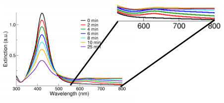
Aggregation of silver nanoparticles increases long-wavelength extinction, including near wavelengths used for kinetic turbidimetric endotoxin analysis.
Endotoxin Binding or Masking
Nanoparticle surfaces can interact with endotoxin. Adsorption of endotoxin to a particle surface can reduce its accessibility to the assay reagent and produce an artificially low measurement.
Porous, hollow, lipid-based, or other complex nanoparticle systems can present additional challenges if endotoxin becomes associated with or entrapped within the formulation.
A normal spike-recovery control demonstrates that externally added endotoxin can be detected under the tested conditions, but it does not necessarily prove that previously entrapped or strongly particle-associated endotoxin is fully recovered. Sample-specific method development may therefore be necessary for complex formulations.
Sample Preparation for Nanoparticle Endotoxin Analysis
Sample preparation should reduce assay interference without changing the amount of endotoxin that the analysis is intended to measure.
Use Endotoxin-Controlled Materials
Use endotoxin-free or appropriately qualified water, tubes, pipette tips, labware, and other materials throughout sample preparation. Because the assay is highly sensitive, environmental or handling contamination can contribute measurable endotoxin.
Powder samples may require dispersion in endotoxin-free water or another qualified medium before testing. The selected preparation should be compatible with both the material and the assay.
Dilution
Dilution is one of the most useful approaches for reducing nanoparticle-related assay interference. It can decrease optical density, particle concentration, ionic strength, and the concentration of interfering formulation components.
However, dilution also raises the effective quantification limit for the original sample. A 100-fold dilution, for example, means that the assay must detect one-hundredth of the original sample concentration to achieve the same reporting limit after dilution correction.
For regulated bacterial endotoxin testing, dilution should not exceed the applicable maximum valid dilution (MVD). Dilution beyond the MVD could reduce an unacceptable endotoxin concentration below the detection capability of the assay.
pH
LAL reactions require an appropriate pH range for normal enzymatic activity. Strongly acidic or basic samples may inhibit the assay.
The LAL reagent itself provides some buffering capacity, but samples with extreme pH may require dilution or pH adjustment using qualified reagents. Any sample treatment should be evaluated to ensure that it does not alter endotoxin recovery.
Separating Nanoparticles from the Sample
For highly interfering nanoparticle formulations, separating the particles before analysis may reduce optical interference. Centrifugation, ultrafiltration, or another appropriate method can sometimes be used to test the particle-depleted fraction.
This approach changes the measurand. Endotoxin associated with or entrapped by nanoparticles may be removed along with the particle fraction, so the result should not automatically be interpreted as the total endotoxin content of the original formulation.
Any separation approach should be evaluated for both nanoparticle removal and endotoxin recovery.
Suggested Starting Dilutions for Concentrated Silver Nanoparticles
The following dilutions were developed historically for 1 mg/mL aqueous silver nanosphere formulations to reduce optical interference during kinetic turbidimetric analysis. They should be considered starting points rather than universal assay conditions.
| Silver Nanosphere Diameter | Suggested Starting Dilution |
|---|---|
| 10 nm | 1:500 |
| 20 nm | 1:250 |
| 30 nm | 1:150 |
| 40 nm | 1:100 |
| 50 nm | 1:100 |
| 60 nm | 1:100 |
| 80 nm | 1:50 |
| 100 nm | 1:50 |
Historical starting dilutions for 1 mg/mL aqueous silver nanosphere formulations using kinetic turbidimetric LAL analysis. Appropriate dilution should be established experimentally for each formulation using assay-interference controls.
Inhibition and Enhancement Controls
A critical part of endotoxin testing is demonstrating that the sample does not prevent the assay from detecting endotoxin or artificially increase the measured response.
An inhibition/enhancement control, also called a positive product control, is prepared by adding a known amount of endotoxin to the sample and measuring how much of that spike is recovered.
Spike recovery can be calculated as:
Spike Recovery (%) = [(Spiked Sample − Unspiked Sample) / Added Endotoxin] × 100
For NCL methods based on USP bacterial endotoxin testing principles:
| Spike Recovery | Interpretation |
|---|---|
| 50–200% | Generally considered acceptable recovery for the inhibition/enhancement control. |
| <50% | Indicates assay inhibition and potential underestimation of endotoxin. |
| >200% | Indicates enhancement, contamination, or another source of an artificially elevated response. |
If interference is detected, the endotoxin result at that sample condition should not be considered valid. Testing at an appropriate additional dilution or using another validated sample-treatment or detection approach may resolve the interference.
Common Sources of Inhibition or Enhancement
Interference can arise from:
- Nanoparticle optical extinction or scattering
- Nanoparticle aggregation during the assay
- Endotoxin adsorption to particle surfaces
- High or low pH
- High concentrations of salts, surfactants, proteins, or other excipients
- Proteases that interact with assay components
- β-glucans that can activate Factor G in conventional LAL reagents
- Other formulation-specific interactions with the assay cascade
β-glucan interference is particularly relevant to conventional LAL assays because some lysates contain a Factor G pathway that responds to β-glucans independently of endotoxin. Glucan-blocking approaches, Factor-C-specific reagents, or recombinant Factor C methods can help distinguish or avoid this interference when appropriate.
Interpreting Endotoxin Results
Endotoxin results should be interpreted together with sample dilution, assay range, interference controls, replicate precision, and the purpose of the measurement.
EU/mL and Dilution Correction
If a sample is diluted before analysis, the concentration measured by the assay must be multiplied by the dilution factor to determine the endotoxin concentration in the original sample.
For example, if a 1:100 dilution measures 0.10 EU/mL:
Original Sample Concentration = 0.10 EU/mL × 100 = 10 EU/mL
Report Values Only Within the Qualified Analytical Range
A result above or below the calibrated assay range should not be treated as equally reliable to a value measured within the qualified range. Samples above the range can generally be diluted further and remeasured, provided the final dilution remains appropriate for the assay.
When endotoxin is below the reporting limit, report the result relative to the method's applicable detection or quantification limit rather than assigning a numerical value by extrapolating beyond the standard curve.
Endotoxin Limits Depend on the Application
There is no universal acceptable endotoxin concentration for every nanoparticle formulation. Appropriate limits depend on factors such as intended use, route of administration, dose, experimental model, and applicable regulatory or compendial requirements.
For research samples, the practical question may be whether endotoxin is low enough to avoid confounding a particular biological experiment. For regulated pharmaceutical or medical-device applications, acceptance criteria should be established from the applicable product-specific and regulatory requirements.
What to Report with Endotoxin Data
For interpretable and reproducible endotoxin analysis, report relevant information such as:
- Endotoxin test format and reagent type
- Sample matrix and nanoparticle formulation
- Sample dilution factor
- Measured and dilution-corrected endotoxin concentration
- Assay calibration range
- Positive product control or inhibition/enhancement recovery
- Replicate precision when applicable
- Relevant pH or optical-interference considerations
- Any filtration, centrifugation, neutralization, or other sample treatment
- Applicable detection or quantification limit
Troubleshooting Nanoparticle Endotoxin Analysis
| Observation | Possible Causes and Next Steps |
|---|---|
| High baseline optical density | The nanoparticle sample may absorb or scatter strongly at the assay wavelength. Evaluate an appropriate dilution or alternative assay format. |
| Optical density increases before the expected LAL response | Nanoparticles may be aggregating after addition of assay reagents. Monitor particle stability and test an appropriate lower particle concentration. |
| Spike recovery <50% | The formulation is inhibiting endotoxin detection. Evaluate additional dilution within the allowed range or another qualified interference-control strategy. |
| Spike recovery >200% | Consider assay enhancement, contamination, β-glucan interference, or another sample-specific interaction. |
| Replicate results show poor precision | Check sample homogeneity, optical interference, aggregation, pipetting, assay timing, and contamination. |
| Result decreases substantially with dilution | Evaluate whether the assay is experiencing enhancement at lower dilution or whether sample handling is altering endotoxin recovery. |
| Result increases after dilution | The concentrated formulation may be inhibiting endotoxin detection. Review spike recovery at each dilution. |
| Chromogenic assay gives inconsistent results for a colored nanoparticle | Particle extinction may overlap the assay wavelength. Consider a different assay format or demonstrate adequate optical correction and spike recovery. |
| Particle-depleted supernatant has little endotoxin | Endotoxin may be associated with the particles or lost during separation. Confirm recovery before interpreting the supernatant as total endotoxin. |
| LAL result appears elevated in a β-glucan-containing formulation | Factor G activation may contribute to the signal. Consider a glucan-specific control or a Factor-C-specific/recombinant method where appropriate. |
The most important question is not simply whether the instrument produces an EU/mL value, but whether the assay has demonstrated that it can accurately recover endotoxin from the specific nanoparticle formulation being tested.
Have questions about endotoxin analysis for your nanoparticle formulation?
Talk with our technical team about sample preparation, nanoparticle interference, dilution strategy, assay controls, or endotoxin testing for your material.
Selected References
- Neun, B.W. and Dobrovolskaia, M.A. Detection of Endotoxin in Nano-formulations Using Limulus Amoebocyte Lysate (LAL) Assays. Journal of Visualized Experiments. 2019;(143).
- Neun, B.W. and Dobrovolskaia, M.A. Detection and Quantification of Gram-Negative Bacterial Endotoxin Contamination in Nanoparticle Formulations by Kinetic Turbidity LAL Assay. National Cancer Institute Nanotechnology Characterization Laboratory Assay Cascade Protocols.
- Smulders, S., Kaiser, J.P., Zuin, S., et al. Contamination of nanoparticles by endotoxin: evaluation of different test methods. Particle and Fibre Toxicology. 2012;9:41.
- Dobrovolskaia, M.A. Pre-clinical immunotoxicity studies of nanotechnology-formulated drugs: Challenges, considerations and strategy. Journal of Controlled Release. 2015;220:571-583.
- USP <1085> Guidelines for Bacterial Endotoxins Testing. United States Pharmacopeia, 2025.
- USP <86> Bacterial Endotoxins Test Using Recombinant Reagents. United States Pharmacopeia.
Other nanoparticle characterization guides