
Polyvinylpyrrolidone (PVP) is a polymer commonly used to stabilize metal nanoparticles. At nanoComposix, we typically use 40 kDa PVP, which adsorbs strongly to the nanoparticle surface and creates a steric barrier that helps prevent particles from contacting and aggregating when solution conditions change or when particles are dried.
Compared with smaller, more readily displaced stabilizers such as citrate or tannic acid, PVP is more persistent on the particle surface. It is particularly useful when nanoparticles need broad solvent compatibility, high salt tolerance, or the ability to be dried and subsequently redispersed.

Looking for PVP-stabilized nanoparticles?
Explore PVP-coated gold and silver nanospheres, including dried formats for greater flexibility in solvent selection, or talk with our team about a custom formulation.
Gold Nanospheres • Silver Nanospheres • Custom Nanoparticles
PVP Surface Properties
- Surface character: Sterically stabilizing polymer coating
- Surface charge: PVP itself is nonionic, although PVP-coated metal nanoparticles can exhibit a negative measured zeta potential
- Displaceability: Less readily displaced than citrate, carbonate, or tannic acid
- Salt stability: Very high in the 40 nm gold nanoparticle formulation tested below
- Drying and redispersion: PVP can help particles withstand drying and subsequent redispersion in compatible solvents
- Solvent compatibility: Compatible with water and a range of polar organic solvents, depending on the nanoparticle formulation
Representative source material: Polyvinylpyrrolidone (MilliporeSigma / Calbiochem, 5295)
Representative molecular weight: 40 kDa
Why Use a PVP Surface?
The primary advantage of PVP is its combination of steric stabilization and broad formulation flexibility. The polymer layer physically separates neighboring particles, reducing their tendency to make direct contact even when electrostatic stabilization is weakened.
This makes PVP useful for nanoparticles that may encounter changes in salt concentration, pH, solvent composition, or processing conditions. PVP can also help preserve particle dispersibility when nanoparticles are dried onto substrates, incorporated into thin films, or supplied as dried powders for subsequent redispersion.
PVP can be displaced under some conditions by ligands with strong affinity for noble-metal surfaces, including thiol-containing molecules. However, when efficient ligand exchange is the primary goal, a more accessible surface such as citrate is generally a better starting point.
Applications
- Dry nanoparticle powders and solvent redispersion
- Transfer between compatible aqueous and organic solvents
- Thin films, coatings, and composite materials
- Color engineering and plasmonic materials
- Applications requiring high colloidal stability across changing solution conditions
For optical and materials applications, see Nanomaterials for Optical Engineering.
PVP Surface Charge

The figure above shows representative zeta potential versus pH data for PVP-coated 40 nm gold nanoparticles. These data were generated by manual titration with HCl and NaOH followed by zeta potential measurement.
Although PVP is a nonionic polymer, the complete PVP-coated nanoparticle interface exhibits a negative measured zeta potential across most of the pH range tested. In this dataset, the zeta potential approaches neutral only under strongly acidic conditions, with an apparent crossover below approximately pH 3.
The magnitude of the negative zeta potential increases as pH becomes more basic until approximately pH 10. At higher pH in this titration series, the magnitude begins to decrease, likely because increasing ionic content compresses the electrical double layer.
nanoComposix has observed similar overall pH-dependent zeta-potential behavior between comparable gold and silver nanoparticles for other charged surfaces, including citrate and BPEI. These observations provide qualitative context when comparing metal nanoparticle systems, although the PVP dataset shown here was measured specifically using 40 nm gold nanoparticles.
Learn more about how pH, ionic strength, and particle surface chemistry affect these measurements in Zeta Potential Measurements.
Salt Stability of PVP-Coated Nanoparticles

PVP provides steric stabilization, so colloidal stability does not depend exclusively on electrostatic repulsion. This allows PVP-coated nanoparticles to tolerate much higher ionic-strength conditions than surfaces that rely primarily on charge stabilization.
The figure above shows UV-Visible spectra of PVP-coated 40 nm gold nanoparticles exposed to increasing concentrations of sodium chloride (NaCl).
Stable 40 nm gold nanoparticles retain their characteristic plasmon resonance near 520 nm. Aggregation would typically produce a decrease in the primary plasmon peak and increased extinction at longer wavelengths, approximately 700–1100 nm, due to plasmon coupling between neighboring particles.
In this dataset, the PVP-coated gold nanoparticles remain stable in saturated NaCl solution, with no substantial long-wavelength spectral feature associated with aggregation. This demonstrates the high salt tolerance that a PVP coating can provide under the conditions tested.
This result is specific to the 40 nm gold formulation and experimental conditions. Salt stability can vary with particle material, size, PVP molecular weight, surface coverage, concentration, and formulation. nanoComposix has generally observed lower salt stability for silver nanoparticles than for comparable gold nanoparticles with the same surface chemistry.
See Salt Stability of Nanoparticles for comparisons among different nanoparticle surface chemistries.
Solvent Compatibility & Redispersion
Many PVP-stabilized nanoparticles can be dried and subsequently redispersed in compatible solvents. To redisperse a dried powder, add the selected solvent and bath sonicate for approximately 30 seconds.
The table below summarizes redispersion behavior observed for PVP-coated nanoparticle powders. Compatibility can vary with particle material, size, PVP coverage, concentration, and processing history, so these values should be treated as formulation guidance rather than universal solvent specifications.
| Solvent | Refractive Index, n | Dry Particle Redispersibility |
|---|---|---|
| Water | 1.34 | High |
| Methanol | 1.33 | High |
| Ethanol | 1.36 | High |
| Isopropanol | 1.38 | High |
| DMF | 1.43 | High |
| DMSO | 1.48 | High |
| Chloroform | 1.45 | Medium |
| Acetonitrile | 1.34 | Low |
| Dichloromethane | 1.42 | Low |
| THF | 1.41 | None |
| Hexane | 1.38 | None |
| Toluene | 1.50 | None |
After redispersion, samples should be stored at 4°C and protected from light. See Storage & Handling for additional guidance.
Effect of Solvent on Nanoparticle Optical Properties
Changing the solvent surrounding a plasmonic nanoparticle can shift its optical spectrum even when the particle itself remains unchanged. UV-Visible spectroscopy can therefore be used to evaluate redispersed particles, but spectral shifts must be interpreted in the context of the refractive index of the new solvent.
In the experiments below, PVP-coated silver nanoparticles with diameters of 10, 50, and 100 nm were dried into powder form and redispersed in a variety of solvents. The spectrum of each redispersed sample was compared with that of the original aqueous dispersion.
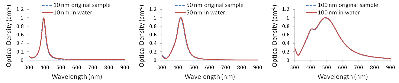
UV-Visible spectra of PVP-coated silver nanopowders redispersed in water (n = 1.33).
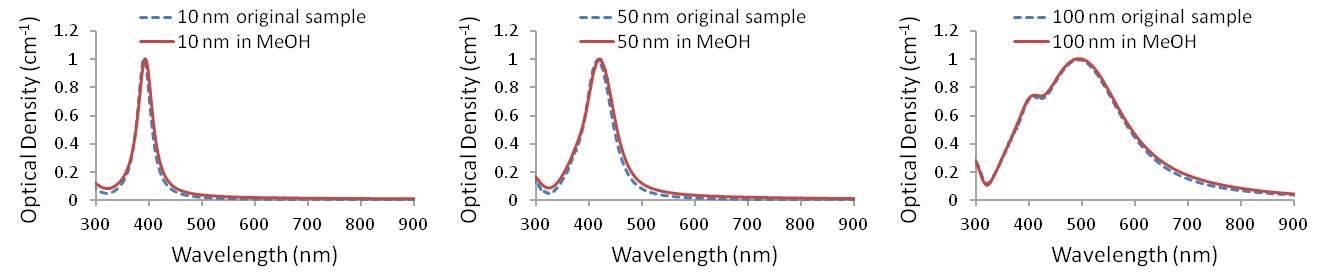
UV-Visible spectra of PVP-coated silver nanopowders redispersed in methanol (n = 1.33).

UV-Visible spectra of PVP-coated silver nanopowders redispersed in ethanol (n = 1.36).
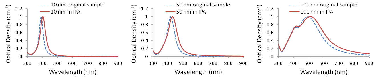
UV-Visible spectra of PVP-coated silver nanopowders redispersed in isopropanol (n = 1.38).

UV-Visible spectra of PVP-coated silver nanopowders redispersed in DMF (n = 1.43).
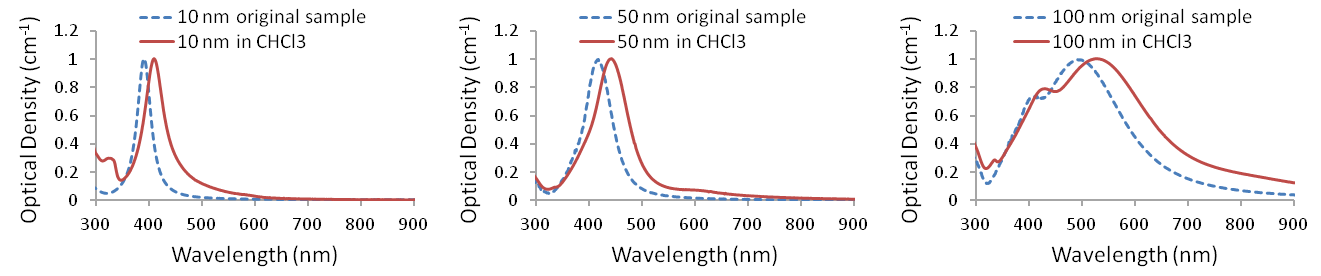
UV-Visible spectra of PVP-coated silver nanopowders redispersed in chloroform (n = 1.45).
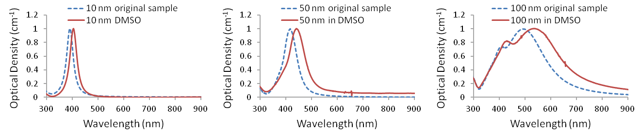
UV-Visible spectra of PVP-coated silver nanopowders redispersed in DMSO (n = 1.48).
Redispersion in water and methanol produces spectra that are nearly indistinguishable from the original aqueous dispersions. Larger spectral differences appear in several of the other solvents, but most of these changes can be explained by the different refractive index of the surrounding medium rather than by nanoparticle aggregation.
For example, Mie theory predicts a shift in the optical spectra of 10, 50, and 100 nm silver nanoparticles when the surrounding refractive index changes from that of water to that of chloroform.

Calculated spectra for silver nanoparticles in water (n = 1.33) and chloroform (n = 1.45). Increasing solvent refractive index shifts and broadens the plasmonic features.
The experimentally observed shifts and broadening agree well with the predicted response. In these datasets, only the small secondary feature near 650 nm for the 50 nm particles and slight broadening of the long-wavelength tail for the 100 nm particles indicate low levels of agglomeration.
More generally, increasing the refractive index of the surrounding solvent shifts the silver nanoparticle plasmon resonance toward longer wavelengths. The comparison below shows the measured peak wavelengths for 10, 50, and 100 nm silver nanoparticles in different solvents alongside values predicted using Mie theory.
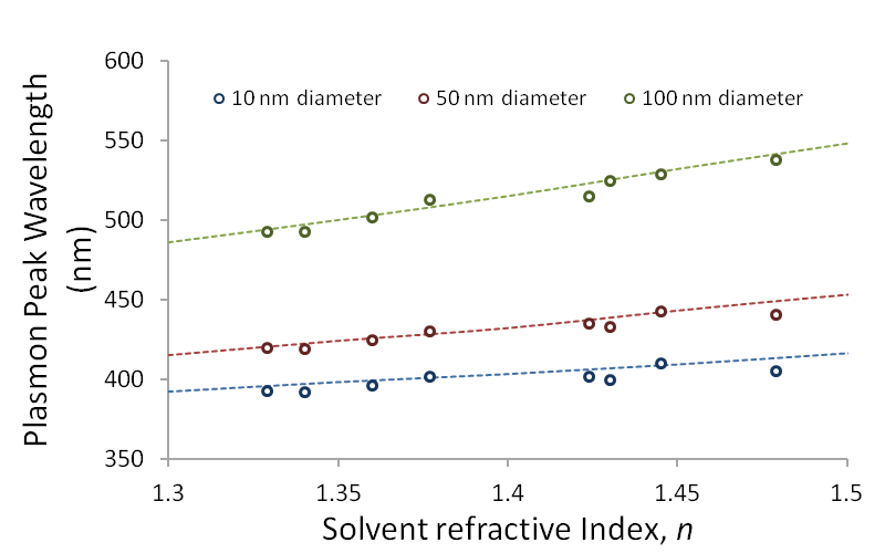
Measured plasmon peak wavelengths for 10, 50, and 100 nm silver nanoparticles in different solvents compared with Mie theory predictions.
Learn more about the effect of particle environment on optical response in Silver Nanoparticle Optical Properties or explore particle and solvent effects using the Mie Theory Calculator.
Effect of Solvent on Hydrodynamic Diameter
Dynamic light scattering (DLS) provides a complementary way to evaluate redispersed nanoparticles. Unlike TEM, which measures the physical dimensions of the metallic particle, DLS measures an apparent hydrodynamic size influenced by the PVP layer, its interaction with the solvent, and the particle's surrounding solvation environment.
The table below shows the average hydrodynamic diameter measured for nominally 50 nm PVP-coated silver nanoparticles after redispersion in different solvents.
| Solvent | Hydrodynamic Diameter (nm) |
|---|---|
| Water | 66.1 |
| Methanol | 69.4 |
| Ethanol | 76.4 |
| Isopropanol | 84.8 |
| DMF | 65.1 |
| DMSO | 71.7 |
The differences among solvents illustrate why DLS measurements should be interpreted in the context of both the nanoparticle coating and the dispersing medium rather than compared directly with TEM diameter as though the two techniques measure the same quantity.
Is PVP the right surface for your nanoparticle system?
Talk with our technical team about solvent compatibility, dried nanoparticle formats, colloidal stability, optical properties, particle size, or selecting a custom surface chemistry.
Related surface chemistry resources